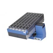

Dual color multichannel fiber photometry system
High accurate
- Ultra-high sensitivity scientific research-grade CMOS camera, with higher quantum conversion efficiency, and the acquisition frequency can reach 300fps.
- Two kinds of excitation light sources (410/470nm), 410nm light source as the reference light source, 470nm light source excites GCaMP, effectively removes motion artifacts.
High efficiency
- Supports up to 9 channels of simultaneous acquisition, suitable for simultaneous recording of multiple nerve nuclei.
- Professional integrated software, fluorescence data and animal behavior video can be collected and analyzed simultaneously.
Humanized
- 4 input ports, support a variety of external signal input and automatic marking, 4 Output ports, support outputting TTL signals to trigger external third-party equipment.
- The software supports 10 kinds of manual marking and automatic marking of external signals.
- The software supports the setting of multiple start and end conditions. And Two modes of excitation Continuous or Sequential.
- Different ROI regions can be set and named during behavioral video collection, and various behavioral analyses can be performed later.
- The results can be exported to CSV, HeatMap, df/f, Z-Score and other formats.
What is Fiber Photometry?
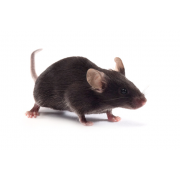
Calcium imaging enables neuroscientists to visualize the activity of large neuronal populations using fluorescent activity indicators (Grienberger & Konnerth, 2012). The invention of imaging technologies, such as the optical fiberscope or miniscope, allows for calcium imaging to be performed in freely-behaving animals with single-cell resolution (Ghosh et al. 2011). These capabilities enable neuroscientists to map the relationship between neural circuits and behavior with single-cell resolution.
Despite many advantages, single-cell resolution calcium imaging systems are (1) more complex and generate large data sets that are difficult to handle, and (2) require more intricate surgical procedures. For labs just starting with calcium imaging or performing exploratory experiments, single-cell imaging may be too overwhelming or unnecessary.
So, is there a SIMPLE and LOW COST technology for in vivo calcium imaging?
As a technique of monitoring the neuronal activity of neurons labeled with a fluorescent reporter(s) in behaving lab animals that uses chronically implanted optical fiber to deliver excitation light and collects induced fluorescence from the targeted brain region, Fiber photometry has become a very popular method for in vivo calcium imaging due to its many advantages. Fiber photometry Researchers can use genetic animal models and fluorescent proteins, such as calcium (GCaMP) and dopamine (dLight), to study specific brain circuits.
Advantages:
- Record neural activity of a specific neuron population
- Compatible with deep brain fiber-optic cannula
- Low impact on an animal in freely behaving experiment
- Allows for multiple sites recordings
- High temporal resolution
- High sensitivity, low optical power required
What components are needed to perform fiber photometry?
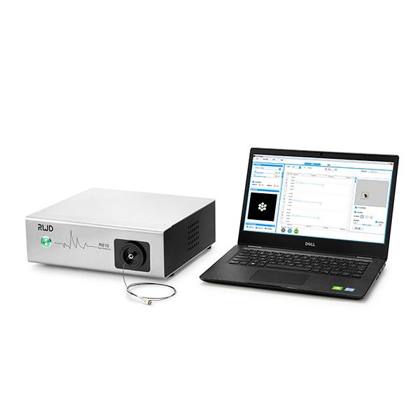
RWD offers complete solutions for fiber photometry going from data acquisition and analysis software, light sources, patch cords, and rotary-joints to fiber-optic cannulas and related accessories. The R810 Fiber Photometry is an out-of-the-box solution designed for researchers. Our user-friendly software provides a seamless interface to the real-time processor and integrated streamline hardware setup and debugging allow you to focus on the research and not the equipment.
Features
- Two excitation ranges centered at 410/470nm, triple color will be available soon.
- Record from multiple locations (9 channels of simultaneous acquisition) in a single animal or many animals simultaneously with a single system
- Integrated digital signal synchronization module, 4 Input and output interfaces respectively, meeting closed-loop research.
- Professional integrated software, fluorescence data and animal behavior video can be collected and analyzed simultaneously.
- Support multiple start and end condition settings and multiple excitation light output modes.
- For complex experiments with multiple stimuli, 10 kinds of manual marking and automatic marking are supported, and 9 behavioral ROIs can be set.
- The analysis results can be exported to CSV, Heat-map, df/f, Z-Score and other chart formats.
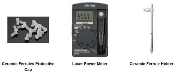
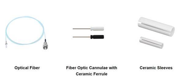
Accessories
Fiber optic cannula (FOC) with black ceramic ferrules that have been optimized for fiber photometry with an 80% efficiency cutoff. Be sure to opt for a low autofluorescence fiber and match the numerical aperture (we can help with this).
- Frame Rate of CMOS - 0~300 FPS
- sCMOS camera resolution - 2048*2048px
- Exposure time setting range - 1~100 ms
- Fluorescence band - 500-550 nm
- Central wavelength of excitation light - 410 nm、470 nm
- LED power adjustable percentage - 0~100%
- 410nm LED Output Power Range - 0~100 μW
- 470nm LED Output Power Range - 0~100 μW